Showing posts with label neat Bacteria. Show all posts

Sunday, April 1, 2012

Apparently, Bacteria have ears...

Bacteria have an endless ability to amaze. Even plain old Escherichia coli, the workhorse organism of thousands of labs—an organism that, in my youth, I thought too boring and well-known to be worth focusing a career on—still generates research that leaves us dumbstruck and groping for explanations. Nowadays, as often as not, these surprises turn up on the fringes of science and don’t really affect our understanding of how the world works. However, they do make us pause and scratch our heads—sometimes at the result itself, other times at why the initial experiments were done. So, we have findings from Czech researchers about how E. coli in “heavy” water (enriched for the deuterium isotope of hydrogen, 2H2O) grow faster than they do in normal water. We have the finding from an astrobiology group that growth in zero gravity causes changes in the expression of almost half of E. coli’s genes. And now, a new report suggests that E. coli can actually sense and respond to sound—in a word, bacteria can hear.


The finding, like many in the history of science, is an example of Pasteur’s dictum about fortune favoring the prepared mind. In this case, the mind belongs to Felix Balatro of Miskatonic University. He was actually testing a pet hypothesis about whether Bacteria could learn.


Of course, one would not expect terribly deep thinking from a bacterium. Nor would you expect deep thinking from the flatworm Planaria—however, this has been demonstrated using a T-maze. A T-maze is the simplest possible maze: the worm can go forward from the bottom of the T to the top, then it can turn left or right. If the reward is consistently on the right, then the little guys start turning right more than half of the time. Balatro had the rather crazy idea that bacteria could do the same trick. One problem he had to solve was a matter of scale: a planarial T-maze is a few centimeters long, a distance that would take a couple of generations for E. coli to swim. Fortunately, advances in microfluidics and techniques borrowed from semiconductor production enabled him to produce a T-maze of millimeter scale, with exactly equal concentrations of an attractant (aspartate) at each branch of the maze.


Now, the first piece of luck for Balatro was that he didn’t really think this through. Planaria, being flatworms, have a clearly defined left and right. They crawl, instead of swimming. So, the right side of the T-maze corresponds only to the right side of the flatworm’s body. Not so with E. coli. They swim, and spiral and loop and frequently tumble as they swim, so they “know not their right hand from their left.” Anyone knowing this basic fact of E. coli’s biology knows that this experiment would fail—but Balatro, either through ignorance or (more likely) stubbornness, pushed ahead.


Amazingly, E. coli seemed to learn to turn right. Balatro ran his cells through the T-maze, and collected the cells that made it to the right side of the maze. Using some nifty microfluidic devices, he let these cells reproduce for one generation, and ran them through the maze again. Again he selected the cells that turned right, and let them grow again…and again, and again, for approximately a thousand generations. After this time, when he ran a thousand cells through the maze, he found that between 60 and 70% of the cells would turn right.


This result is completely, totally, 100% unbelievable. Being a good scientist, Balatro didn’t believe it. Suspecting some barely perceptible turbulence in his test chamber, he turned the microfluidic slide upside down in the microscope—and obtained the same result. He wondered if the bacteria were somehow responding to light, so he blacked out his lab and ran the cells through the maze with the microscope’s lights out—and again found that nearly three quarters of the cells turned right.


Magnetotaxis—the ability to sense and respond to the Earth’s magnetic field—is not unknown among the bacteria, so Balatro tried to test this. “Right” in his original maze corresponded to south-southeast. He spent a day rearranging the tangle of pumps and controllers surrounding the apparatus so that “right” now corresponded to north-northwest, and he must have experienced a mixture of surprise and elation when he found that now, 75% of his cells got to the T and turned left!


Balatro’s celebratory mood was presumably deflated when he shared his results with his colleague, D. Avril Poisson. She looked through the microscope at the maze, coolly removed a magnet from a nearby refrigerator door, and set it upon the stage. The resulting magnetic field was many times stronger than the Earth’s, and the bacteria did not change their behavior at all. In fact, when Balatro ran the experiment in her presence, the bacteria seemed to forget everything—they got to the T, and half turned left, half turned right.


Here’ was Balatro’s second piece of luck. It had to do with a combination of his intellect and his personal eccentricities. I got to know one of Balatro’s PhD committee members when I was in graduate school, and she described him as “one of the most brilliant and…um…uh…ehhh…I guess you could say lateral thinkers I’ve met.” He apparently lived in the lab, only going home to shower; he spent a fair amount of his time in lab stoned, and he spent absolutely all of his time in lab, awake or asleep, with the stereo blasting the Grateful Dead at deafening volume. He even ditched one thesis advisor (and, thus one field of study and one future career) for another solely because his newly chosen advisor was deaf and didn’t mind the noise—and so oncology’s loss was microbiology’s gain.


Rather than mope about a failed experiment, Balatro realized that only one thing had changed when Poisson had entered the room. Out of hard-learned politeness, he had muted Jerry Garcia’s solo from “Sugaree” at the Rockpalast, 1977, blasting from the speakers in the south end of his lab. Nothing else had changed—nothing about light, the magnetic field, temperature, pH, nutrient or redox gradients, or any of the myriad other things bacteria are known to sense. Just pure, Gratefully Dead sound. Had he discovered phonotaxis?


Of course, Balatro confirmed this result, and spent a lot of time designing experiments that eliminated other possibilities and left the conclusion that his bacteria could hear and turn towards sound. And so we have the paper, given the innocuous title “Novel functions for mechanosensitive channel proteins in an evolved Escherichia coli system,” and slipped into print in the obscure journal Acta Pathologica, Microbiologica et Immunologica Scandinavica. It’s as if Balatro doesn’t want us to notice that he has discovered something incredible.


However, this is a powerful paper; Balatro, working with Poisson, even proposes a plausible mechanism. Sound, at the cellular level, is a mechanical phenomenon—an alternation of high and low pressure. Bacterial cells have mechanosensory proteins in their membranes, and these have been intensively studied for their role in helping cells survive osmotic stresses. These proteins, as they sit in the cell’s membrane, are deformed when the cell membrane is deformed; the change in the protein’s shape causes the protein to send a signal into the cell, resulting in a change in the expression of several genes.


Balatro and Poisson examined the genes for these mechanosensory proteins, mscL and mscS, in the cells that consistently turn towards high-intensity sound, and found that these genes had evolved over the course of generations of selection. The genes had, in fact, recombined with genes for chemotaxis—that is, genes that encoded proteins that sensed food, and directed cells to swim towards higher concentrations of food. The evolved gene encoded a hybrid protein that still sensed mechanical pressure (though this too had changed slightly), but now controlled the cell’s swimming behavior.


This result is curious, and leaves open lots of questions. We still don’t quite understand how sound waves, which should be much larger than a bacterial cell, can have a perceived direction to E. coli—it would be as if we could tell, just using our own senses, what direction a change in barometric pressure came from. There is also the puzzle of how the changes in the pressure-sensing component of the MscL and MscS proteins in these “hearing” bacteria relate to their ability to hear. Information about the structure of these mutant proteins should provide some answers, and Balatro is quite able to get that data rapidly.


Ultimately, like cells growing in Deuterium or microgravity, this is a completely artificial situation, and thus not likely to be of especial biological significance. Still, it’s pretty neat, and a good reminder of both how science progresses and how amazingly extraordinary even “ordinary” bacteria can be.


Mann, L.R., and Moses, V. (1971). Properties of Escherichia coli grown in deuterated media. Folia Microbiol Praha 16 (4): 267-84.


Rosenzweig, J.A., Abogunde, O., Thomas, K., Lawal A., Nguyen, Y-U., Sodipe, A., and Jejelowo, O (2010). Spaceflight and modeled microgravity effects on microbial growth and virulence. Applied Microbiology and Biotechnology 85 (4): 885-891.


Jacobson, A.L, Fried, C., and Horowitz, S.D. (1966) Planarians and memory. Nature 209: 599-601.


Matsunaga, T., and Okamura, Y (2003). Genes and Proteins involved in Bacterial Magnetic Particle Formation. Trends in Microbiology 11(11): 536-41.


Balatro, Felix, and Poisson, D. Avril (2012). Novel functions for mechanosensitive channel proteins in an evolved Escherichia coli system. Acta Pathologica, Microbiologica et Immunologica Scandinavica 120 (4): 4652-4661.


Haswell, Elizabeth, Phillips, R., and Rees, D.C. (2011) Mechanosensitive Channels: What Can They Do and How Do They Do It? Structure 19: 1356-1369.

Tuesday, March 20, 2012

A lesson from Botox

There's not much in common between what the Real Doctor studies--ophthalmology--and my chosen field of microbial genetics and physiology. One of these rare commonalities is an interest in Botox. For the Real Doctor, it's a handy tool to paralyze an ocular muscle and cure a case of walleye. For me, "Botox" is botulinum toxin, a poison secreted by Clostridium botulinum, and a nice example of how bacterial toxins work.


Right off the bat, I gotta say that bacterial toxins are amazing. It always gives me pause when I see examples of evolution reaching across domains of life. I mean, it is not too surprising when a bacterium evolves a chemical signal to communicate with another bacterium--after all, they share the same biochemistry. But with toxins, bacteria have evolved a very complicated set of molecules that target very specific proteins on the surface of very specific nerve cells in only a few types of organisms; the bacterium is reaching across domains to communicate biochemically with an alien biochemistry.


That said, there are other reasons to be interested in bacterial toxins. They’re amazingly potent—30 nanograms of botox, or a volume of about one billionth of a sugar cube, is enough to kill a human. Their power and specificity makes them valuable tools for understanding the biochemical processes of our own cells, and some (such as botox) have medical uses. Bacteria, in their diversity, have evolved a huge number of bacterial toxins specific to different tissues in different hosts, but most of these toxins are variations on a couple of themes.


One major class of bacterial toxins can be thought of like nuclear missiles; they require two sophisticated components to do their deadly job. By itself, an A-bomb is not so useful a weapon--unless you deliver it to its target, you will only damage yourself. By itself, a guided missile won't do too much damage to its target--it's merely a bus to deliver the payload. But put together the A-bomb and the bus, and you have yourself a tremendously destructive weapon. The bacterial "A-B" toxins are the same way. The "A" component of these toxins is like the A-bomb--a tremendously destructive protein molecule, but it needs to be delivered to its target. The "B" component is like the missile (or bus), a protein that is by itself harmless, but protects the "A" component and delivers it to its target.


Botulinum toxin is an example of an A-B toxin, with a wicked but delicate A component and a very interesting B component. The bacteria release this toxin in our guts, an extremely acidic environment, and it must travel into our blood and then find a specific molecule on a specific type of nerve cell to do its dirty work. Some recent work by a group at the Sanford-Burnham Medical Research Institute in La Jolla, California, has given us a peek into how the B component delivers the destructive A component to its target, and also--like any good study--raised some more interesting questions.


The researchers focused on the first stage of this deadly missile's trajectory--the trip from the acidic digestive system into the more neutral bloodstream, a hazardous journey that almost no proteins can survive. As long as people have known that botulinum toxin is made of protein, it has been a mystery how it avoided destruction. So, what about B allowed this? By making various modified versions of the B component, they found that only one of the four parts of this carrier was necessary for protectin the A component from acid. Then, since the function of any protein is intimately tied to its structure, they tried to find out the structure of this minimal B and how it fit together with the A. They found a couple of interesting surprises.


First, the B protein is sensitive to acid, just as susceptible to acid as the delicate A component of the toxin. Both B and A, individually, are attacked by acid at a few specific sensitive places, and break into a few distinctive pieces. However, if you put them together in an acidic environment, they laugh it off. The reason is in how these proteins fit together: all the bits that are sensitive to acid are covered up by the way the proteins nestle up against each other. They snuggle against each other to protect each other from harm, a romantic image if we weren’t dealing with deadly poisons.


A second interesting feature came to light when the researchers looked at exactly how these proteins fit together. The researchers were able to recognize the specific parts each protein that touched each other. These parts were notable because of how they responded to acidity: in the harsh of environment of the stomach, these parts of the proteins remained neutral, and helped to hold the A and B complex together. However, when the protein complex was moved to a neutral environment, these parts of the protein turned acidic--enough that they would cut the bonds between the A and the B proteins. The connections between the A and B proteins acted like the explosive bolts that hold together the stages of a missile, holding them together through the boost phase but dramatically separating the two when the booster is done. Indeed, the researchers conclude that this is how the first part of botulinum toxin's journey goes: the minimal part of the B component protects the A component in the stomach and carries it into the bloodstream. Once the two are in the bloodstream, the neutral environment causes B to cut its links to A, and the A component can go on its merry way.


This is all pretty cool. It solves a mystery about how botulinum toxin works, and actually suggest as way that biotechnologists could design protein-based drugs that could be taken orally. If a protein drug could be designed to fit with a carrier similar to botulinum toxin B component, then it could survive the trip through the stomach and be absorbed into the bloodstream, where it could do its job.


However, for my money, the most surprising (and instructive) thing revealed by the structure of the minimal botulinum toxin is that the "A" and "B" components have almost identical structures. Proteins are linear strings of hundreds of amino acids, and these strings clump and fold together in unique and characteristic ways. The structures are often quite complex, with enough helixes and turns and twists that a single protein's structure would resemble a plate of spaghetti, the the entire platefull were one loooong noodle. It is this unique molecular shape that gives each different type of protein the ability to do its specific job--say, disable a nerve cell for the A component of botulinum toxin, or protect another protein from acid for the B component. It is surprising that two proteins that you might expect to have structures as different as an A-bomb and a booster rocket to look almost the same.


Check out this picture: the A component is an orange stringy ribbon, and the B component is a green stringy ribbon. The two structures are superimposed upon each other, and you can see that they are almost identical. (This is just the first third of each of the proteins, but the remaining 2/3 are available, in stereo, at http://www.sciencemag.org/content/335/6071/977/suppl/DC1)


One of the great big hairy problems of modern biology is how proteins--linear molecules--fold up into unique three-dimensional shapes, and botulinum toxin gives us another bracing surprise here. I teach my intro bio students that the "primary structure," or linear arrangement of amino acids in the protein, determines the three-dimensional structure of the protein. A change in the amino acid sequence should result in a change in the three-dimensional structure. I'd expect the botulinum toxin A and B components to have very similar amino acid sequences, given their structural similarity. However, their amino acid sequences are only 20% identical. That's crazy.


The authors, being most interested in biochemistry, don't pursue a Big Question raised by this--how does this evolve? The genes for botulinum toxin A and B components are probably also about 20% identical (mea culpa--I don't have my computer with me, so I have a hard time looking this up. It is left as an exercise for the interested reader). It's possible that two entirely different genes evolved to have such similar structures, but I don't think it's at all likely. More likely, it is a case of an ancestral gene being duplicated, and then each of the two copies undergoing evolutionary change (this hypothesis is weakly supported by the observation that the A and B genes tend to be clustered). These changes altered the sequences of the proteins, while preserving their three-dimensional structures. This would be like sitting down with a sonnet and a thesaurus, and changing 8 of every 10 words. If you were careful, you could preserve the structure, meter, and rhyme scheme of the sonnet, while completely changing the meaning of the poem—and a textual analysis would show no relationship between the source and the end product.


If I could tell these researchers what to do, I would have them gather more structure and sequence information from a variety of related toxin proteins. It would be fun to make a family tree, and see just how much structure could be conserved as amino acid sequence changes.


Shenyan Gu, Sophie Rumpel, Jie Zhou, Jasmin Strotmeier, Hans Bigalke, Kay Perry, Charles B. Shoemaker, Andreas Rummel, Rongsheng Jin (2012). Botulinum Neurotoxin Is Shielded by NTNHA in an Interlocked Complex. Science 335: 977-981.


Peck, MW (2009). Biology and genomic analysis of Clostridium botulinum. Advances in Microbial Physiology 55: 183-265.

Wednesday, August 24, 2011

;-) ;-) (wink, wink)


No living thing is in equilibrium with its environment. I mean this in a physical-chemical sense; ecologists will discuss how ecosystems are at equilibrium, meaning that populations stay constant. However, to a biochemist, equilibrium is the same thing as death. A system at equilibrium has zero energy, and since life by any definition requires energy, equilibrium is death. It follows that if you can measure how far out of equilibrium a living thing is, you can get a clue as to how much energy it has at its disposal.


The way almost all living things get energy is sort of like the way energy is stored in a battery:

In a battery, there’s too many electrons over at one end, and not enough at the other. This potential difference is a form of energy. We can let the battery get to equilibrium by letting the charges become uniformly distributed, and we can use that movement to do some work for us:

If it achieves equilibrium—an equal concentration of charges on either end—no more work can be done.

A living cell functions almost exactly the same way. The only changes are that the charge difference is between inside and outside, and the charged particles are (positively-charged) protons (H+) instead of (negatively charged) electrons.

And, just like a battery, this potential difference stores energy. If we let the cell get to equilibrium by letting the charges become uniformly distributed, the cell can use that movement to do some work. Instead of powering light bulbs, cells have enzymes in their membranes that use that energy to make a chemical form of energy, ATP, or wiggle their flagella around so they can move, or pack food into the cell and expel wastes.

Also like a battery, equilibrium means no more work can be done.


Establishing the disequilibrium in a battery takes work, and making the proton gradient across a cell membrane also takes work. Respiring cells use the oxidation of food to move protons out of the cell. Cells that can use light for energy have evolved a couple of mechanisms to move protons out of the cell. The simplest of these is called proteorhodopsin. It’s an enzyme that straddles the cell membrane, like so:

When it’s hit by light...

it can use that energy to change its chape, grab a proton from inside the cell and force it out of the cell (and away from equilibrium). Like so:

To understand the physiology of a cell, it’s nice to know the charge difference, or membrane potential, of the cell. Keeping track of membrane potential is pretty straightforward in cells like ours. Our cells are large enough that we can make tools that can investigate to our cell membranes the way that a stethoscope can check in on a guinea pig. However, bacterial cells are much too small for this approach, and their membranes are covered up by cell walls. So until recently, we haven’t been able to probe the membrane potential of individual bacterial cells, despite knowing that membrane potential is essential for the well-being of the cell.


A bunch of researchers at Harvard, led by Adam Cohen, have devised (and are in the process of patenting) and exceedingly clever way to look at membrane potential in single bacterial cells. Remember proteorhodopsin, which absorbs light energy and uses it to pull a proton out of the cell? The reason it’s able to absorb light energy is that it has a color. Grabbing onto a proton changes the energy state of the molecule, so it changes its color a little bit. So, Cohen and his gang figured out a way to modify proteorhodopsin, essentially reversing its normal behavior: if a proton were forced onto this modified enzyme, it would not only change color, it would fluoresce. How can a proton be forced onto this modified proteorhodopsin? Simply have a higher-than-normal concentration of protons inside the cell. So, here's a normal cell, and the engineered proteorhodpsin is dark:

And here's a depolarized cell. The relatively high concentration of protons inside the cell forces one onto the engineered bacteriorhodopsin, and it fluoresces:

Bear in mind that what triggers the light is not the concentration of protons, just the difference in concentration across the cell membrane.

This designed membrane protein doesn’t help the cell out any—it can’t do what normal proteorhodopsin does—but it does serve as a sensitive monitor of the membrane potential of a single bacterial cell. It is, in effect, a very sensitive tool. Cohen’s group embedded single bacterial cells in a gel matrix, hooked one end of the gel matrix up to the positive end of a battery and the other end up to the negative end. The electrical field pushed all of the protons in the matrix towards one end of the cell, depolarizing the other end. Sure enough, that end of the cell lit up like a Christmas tree. Here it is in cartoon form...

and the actual microscope view, as they flipped the polarity from one end of the cell to the other:


So far, this is just a cute tool. Unlike our cells, which are so large that small patches of the membrane can depolarize, bacterial cells are so large that they essentially can’t have any regionalized depolarization under normal conditions. Also, this modified enzyme only lights up when there’s essentially no difference in charge across the cell’s membrane—which condition, if sustained, is the same thing as death. So, what’s the point?


Here’s where this gets interesting, and actually tells us something new and unexpected about how bacterial cells live. When Cohen looked at a bunch of these cells under the microscope, he saw them “blink” at him at irregular intervals. Whatever the reason, something about the cells’ membrane potential would periodically just give out. (This movie is sped up four times actual speed.)

The modified bacteriorhodopsin essentially senses the difference in proton concentration across the cell membrane. So, there’s a couple ways that a blink could happen—either too many protons inside the cell, or not enough protons outside the cell. “Concentration of protons” is simply a long way to say “pH,” so to test this possibility, Cohen loaded his cells with a pH indicator (like Litmus paper, only in convenient liquid form!). When a cell “blinked,” its internal pH stayed the same.


The cell uses this proton concentration difference to do stuff—make ATP, and rotate its flagella, among other things. It’s kind of a rude trick, but if you glue a cell’s flagellum down, the whole cell rotates…

…as long as there is a proton gradient. (By the way, that movie is actually slowed down three times!) When Cohen’s group examined a cell that was “blinking,” they found that the blink correlated with the cell’s rotation slowing or stopping. Since the concentration of protons inside the cell is remaining constant, the concentration of protons outside the cell must drop during a blink. (I’m not going to include a movie of a cell…not moving.)


So why would a cell blink? Why would it periodically deprive itself of energy? There’s one thing that it’s much easier for a cell to do if it no longer has a membrane potential, and that is push positively charged substances out of the cell. There are some toxins and metals that are harmful to the cell that are positively charged, and cells are known to spit these out of the cell under stressful conditions. So, perhaps this is happening during a blink: the cell deliberately depolarizes, and purges itself of toxins. Cohen tried feeding his cells a toxic dye, and sure enough, it could be observed flowing out of the cell only during a blink.


So, this Cohen’s cleverly modified protein ends up being more than just an example of clever engineering; it tells us something new about how cells work. Until very recently, the only way we could study the physiology of bacterial cells was by studying the average properties of billions of cells all at once. Very useful information can be gained that way (just ask the US Census Bureau), but vital information about how individuals work is lost. This modified proteorhodpsin is one of several neat tools that have emerged in the last decade that allow us to study single cells or even single proteins. Just as doing interviews with individuals would change your views of census data, these new tools are changing how we view the lives of bacteria.


Joel M. Kralj, Daniel R. Hochbaum, Adam D. Douglass, and Adam E. Cohen (2011). Electrical Spiking in Escherichia coli Probed with a Fluorescent Voltage-Indicating Protein. Science 333: 345-348.


Thursday, March 24, 2011

Oozing along with Beggiatoa (part II)

(See part I).
So, as my student asked, how do they do that--oozing along so hypnotically?

We don’t know. That’s the bottom line. Beggiatoa is really common, but it turns out to be kind of difficult to study in the lab—traditional laboratory work requires getting a pure culture of nothing but Bedgy, and that seems to be pretty near impossible. So it’s been difficult to answer even fairly basic questions about this guy.


We do have a bit of a clue, courtesy of another creature from the same mud sample:

It’s a cyanobacterium, a type of bacterium that does the traditional kind of photosynthesis. It kind of looks similar, and it oozes along in the same way. You can even see that it sort of spirals along like Bedgy does. Unlike Bedgy, you can grow cyanobacteria in a pure culture in the lab, and study how they do stuff—like move along oozily.


We still don’t know exactly how this cyanobacterium (and Bedgy) moves, but at least we have some clues. One set of clues comes from Egbert Hoiczyk and his coworkers, who propose what they call the “slime-extrusion” model. Each cell in that long chain of cells is girdled with a ring of pores, which they think are "slime jets". Each cell produces a lot of slime and pumps it out of the pores. Outside of the cell, the slime picks up water and expands dramatically, like a dried sponge taking up water. The expanding slime pushes the cell along.


This model has some appeal; Hoiczyk did some impressive microscopy examining these slime jets. They’re distinctive protein structures that are neatly positioned to connect the inside of the cell where slime is made with the outside world. They also note a correlation between the presence of slime jets and the ability to glide in other types of cyanobacteria (alas, they did not check on Bedgy).


There’s another line of evidence supporting the slime-extrusion model, and that is actual evidence that slime is extruded. Hoiczyk simply put India ink (a suspension of very fine, black particles) in the mix with his cyanobacteri, and watched them go:

You can see streamers of ink-stained snot trailing off of the chains of cyanobacterial cells. But this isn’t proof that jets of slime are pushing these cells along—it could be these are streamers of snot trailing after these cells as they move along by some other means. If the model is wrong, thinking that the slime is pushing the cells along could be like thinking that the plume of exhaust is pushing a car along.


An alternative to the slime-extrusion model is raised by a group led by David Adams. According to this “peristaltic” model, the slime is mainly a lubricant that helps the cell move. The work is done by fibers that lie just beneath the cells’ outer membranes; these fibers contract in waves that move along the length of the cell. These fibers have been chemically characterized, and also nicely examined by electron microscopy. Nicely, the fibers are all aligned in a lengthwise spiral, causing the cells to spiral along as they glide.


We still don't know whether either of these models really describes how cyanobacteria move, let alone whether or not that has anything to do with the way that Bedgy moves. It would be nice to do some experiments to tinker with the slime jets and see if that affects motility. It would be nice to see whether or not Bedgy even has slime jets. The best we can say is that the way Bedgy moves looks like the way these cyanobacteria move, and that Bedgy has some genes for slime synthesis that kinda, sorta look like the cyanobacterial genes.

For now, though, we can dip into almost any pond, find this beautiful pearl necklace, and just sit back and wonder.


Egbert Hoiczyk and Wolfgang Baumeister (1998). The junctional pore complex, a prokaryotic secretion organelle, is the molecular motor underlying gliding motility in cyanobacteria. Current Biology 8:1161–1168. Electron micrographs of slime jets, ink-stains of slime, and circumstantial evidence for the slime extrusion model.


Marc Mußmann, Fen Z. Hu, Michael Richter, Dirk de Beer, Andre Preisler, Bo B. Jørgensen, Marcel Huntemann, Frank Oliver Glockner, Rudolf Amann, Werner J. H. Koopman, Roger S. Lasken, Benjamin Janto, Justin Hogg, Paul Stoodley, Robert Boissy, Garth D. Ehrlich (2007). Insights into the Genome of Large Sulfur Bacteria Revealed by Analysis of Single Filaments. PLOS Biology 5 (9): e230. The suggestion that Beggiatoa has slime-producing genes similar to gliding Cyanobacteria.


Nicholas Read, Simon Connell, and David G. Adams (2007). Nanoscale Visualization of a Fibrillar Array in the Cell Wall of Filamentous Cyanobacteria and Its Implications for
Gliding Motility. Journal of Bacteriology 189 (20): 7361-7366. Striking images of the fibers that support the peristalsis model of cyanobacterial motility.


(The literature on this subject is pretty thin, and the dates are old; it's obviously not a subject that attracts much interest, cool as it is.)

Wednesday, March 23, 2011

Oozing along with Beggiatoa (part I)


This beauty is from some mud I looked at in the recently-finished microbial diversity lab.

It's Beggiatoa (Bedgy-a-toe-uh, “Bedgy” to its friends), a pretty common resident of the top layer of muddy sediments; it likes a little oxygen, but not very much. Bedgy oozes up and down in the sediments, between oxygen-rich water and anoxic mud to get oxygen levels juuuuuust right.


Bedgy is easy to find, as common as mud, and absolutely hypnotic. Here’s another specimen under higher magnification, looking like a jeweled necklace. I sat and watched this string of cells oozing by for a few minutes minutes, and I never saw the beginning or end of it. You MUST watch it full-screen.


Your high-school biology textbook probably had a general equation for photosynthesis that went like so:


H2O + CO2 --> C6H12O6 + O2


The atoms in water and carbon dioxide are rearranged to produce glucose and oxygen. This takes a lot of energy, and the energy comes from light.


Bedgy is an important bacterium in the history of science. It was the first organism that was observed to perform “chemosynthesis,” rather than photosynthesis. If you look at the periodic table, you’ll see that Sulfur is in the same column as Oxygen, and that Sulfur behaves chemically in some of the same ways as Oxygen. What’s kind of neat is that Bedgy does this:


H2S + CO2 --> C6H12O6 + S (and some SO4)


A difference between Oxygen and Sulfur is that Oxygen is soluble—it dissolves in water, and floats away in our atmosphere when it’s made by photosynthesis. However, the sulfur that Bedgy produces isn’t soluble—so it forms little semi-solid pellets, and that’s what those little yellow dots are. And unlike photosynthesis, the energy for what Bedgy is doing comes from respiration, not light—so, “chemosynthesis,” not photosynthesis. That same high-school biology textbook with the equation about photosynthesis also says that all life ultimately depends upon energy from the sun; Bedgy is there, oozing along and proving them wrong.


When one of my students looked through the 'scope and saw this, the reaction was "WOW!" followed immediately by "Wait--how does it do that?"


Stay tuned.

Wednesday, March 2, 2011

Bold Traveler

One of my favorite television series is Life on Earth, produced by David Attenborough. Every episode seemed to feature him walking across one of the Dry Valleys of Antarctica, or the Atacama desert, or an iceberg, or solfatara. After noting how hostile the area was, Attenborough would turn over a rock and point at a green smudge or a tiny insect, and in his plummy British accent say “And yet…even here...in this harsh environment, life persists!” For champions of endurance, though, Attenborough should have pointed to microbes. As a harsh environment, it’s hard to beat hot rocks nearly three kilometers underground—but microbial life doesn’t just persist there, it seems to thrive.

Interest in life deep beneath the Earth’s surface has been growing in recent years. There was considerable surprise when deep-drilling projects in the 1980’s revealed that oceanic basalts held diverse communities of Bacteria and Archaea. Because of the way these basalts form, we could figure out how old they were. Amazingly, rock that had been buried for millions of years carried millions and sometimes billions of microbial cells. Microscopic analysis using dyes that only stain living cells showed that the majority of these cells were alive, although living veeeery slowly. These observations stimulated further questions—in fact, the two basic questions asked by all environmental microbiologists: who are these guys, and what are they doing?

“Who is there” is easily answered in this day and age by DNA sequencing. Since Bacteria and Archaea are generally identified on the basis of ribosomal RNA sequence, DNA was extracted from these rock samples and probed for the genes encoding ribosomal RNA. These genes could be sequenced and the results would show “who” lived in young (only tens of thousands of years old) oceanic basalts. The answer is somewhat surprising. The Archaea generally have a reputation as “extremophiles,” capable of living in the harshest environments. They also are about half of the organisms found in the deepest oceans. However, in young oceanic basalts, they are only 10% of a surprisingly diverse population. In fact, there seems to be twice the diversity of bacterial species in the young basalts than in the surrounding seawater.

Probing of older basalts showed that as the environment got harsher, diversity decreased. Drilling 1600 meters beneath the ocean floor (into rocks that were deposited over 100 million years ago), researchers found an environment with temperatures ranging from 60 to 90 degrees Celsius, but with a million cells per gram of rock. These cells were definitely alive. However, unlike the younger basalts, the diversity of life sustained by these deep rocks was very low. Generally, only a few types were found, and they were evenly divided between anaerobic Bacteria and methanogenic Archaea. So there seemed to be a trend: deeper and older meant fewer cells and less diversity.

The current record for human curiosity in this subject was established in 2008. In this case, curiosity was helped by greed, as the scientists gained access to a South African gold mine and sampled the water percolating through porous rocks 2,800 meters below the surface of the earth. Conditions there are beastly—a temperature of 60°C (140°F—give a thought to the humans who work there!). Worse, there’s very little energy available. We humans get energy by burning carbon compounds in oxygen; the bacteria living in ocean sediments get energy by burning carbon compounds in sulfates. But in these deep rocks, it seems at first glance that there’s nothing to burn.

“And yet, even here in this harsh environment, life persists!” Not much life, though, and no diversity. The researchers didn’t actually observe any cells—they simply collected DNA. Although this DNA was somewhat similar to that of known types of bacteria, it was unique—enough so that the researchers postulated that it belong to a previously unknown organism, which they named Desulforudis audaxviator. Amazingly, there is no diversity in this environment. They sampled half a million pieces of DNA from this environment, and 99.96% of it came from D. audaxviator. They found essentially no other DNA in their samples. It appears that D. audaxviator—it means “bold traveler”—lives alone in the rocky depths.

We are used to a “web of life,” with organisms interconnected by chemistry and energy: up here, the plants take light and carbon dioxide to make glucose, while we eat the plants to make carbon dioxide, and so on. There are similar cycles for nitrogen, sulfur and other elements, and ultimately, all of these cycles are powered by energy from the sun. Bold traveler lives by itself, depending on no one and feeding no one, a situation unique in my experience in biology. The researchers’ analysis of Bold traveler’s genes show how it can be a one-species ecosystem. It’s able to take in carbon dioxide and make sugar (instead of photosynthesis, it uses chemosynthesis, a light-independent process). It can also “burn” that sugar, using sulfate instead of oxygen, and get energy. It can fix nitrogen, the same way that the bacteria that live in bean plants do. It contains all the element cycles in itself.

If a solo ecosystem is surprising, the energy that probably powers this ecosystem is amazing. We can tell by its genes that Bold traveler burns its fuel using sulfate in place of oxygen—it has the genes for it. But what is less clear is the nature of the fuel it uses. The fuel may be sugar, but there’s very little of that available in the deep rocks. Its genes suggest that Bold traveler burns hydrogen as its fuel. You might reasonably expect hydrogen to be rare in these rocks—it’s a light gas, and would want to be up in the air. But there is water in these rocks, and there is another critical thing: energy.

You may have seen an apparatus that uses electricity to break water into its constituents: electrodes are dipped into water, and a fairly powerful voltage results in oxygen coming off of one electrode, hydrogen off the other. There are no electrodes in the rocks deep underground, but there is energy: the radioactive decay of elements such as thorium and uranium is able to pry hydrogen away from oxygen. Although we don’t know this for sure, we can calculate that the amount of radioactivity present in these rocks is sufficient to keep cells such as Bold traveler alive. So, our best guess is that Bold traveler is, indirectly, nuclear powered: radioactive decay produces hydrogen from water, and Bold traveler burns that hydrogen using sulfate in the rocks it lives in. Unlike just about every other organism we’ve ever seen, Bold traveler is completely independent of the sun.

This all sounds freaky, and for one trained in the biology of “normal” organisms, rather suspect. But what is normal? If normal is what the majority does, that Bold traveler may be normal, and we are the freaks. All the life we are used to—all the birds and trees and people and whales, all the microbes that live in the soil and oceans—are just the thinnest of films on the surface of the earth. The biosphere we know is at best a few meters thick on a globe twelve and a half million meters across. Sure, the life we know may grow to great densities in this environment, but it is a tiny environment. Now consider bold traveler and its fellows: their environment is poor, but thousands of times larger than ours. A simple calculation shows that the majority of microbial life--perhaps the majority of all life--persists in this harsh environment. It's enough to make me envision a microbial Attenborough, trudging through a lush rainforest or fertile farmland, saying "...and yet, even in this harsh environment, life persists!"


By the way, that name—D. audaxviator—is kind of a joke. It refers to Jules Vernes Journey to the Center of the Earth. The heroes of the story are guided by instructions, in Latin, addressed to the “bold traveler”, or audax viator.

Dylan Chivian et al. (2008). Environmental Genomics Reveals a Single-Species Ecosystem Deep Within the Earth. Science 322: 275-278.

Erwin Roussel et al. (2008). Extending the Sub-Sea-Floor Biosphere. Science 320: 1046.

Cara M. Santelli et al (2008). Abundance and Diversity of Microbial Life in Ocean Crust. Nature 453: 653-657.

Wednesday, February 23, 2011

Caulobacter Crazy (Anti-) Glue

There are few things as annoying as out-of-control glue. Klister is one example—it gets everywhere. In violin-building, I use hide glue which is difficult to control for another reason: it is only sticky for a few minutes, which gives you very little time to get your work done. Ideally, glue should only stick when and where you want it.


The wonderful glue used by Caulobacter is amazing not only for its strength, but as we’ve recently found, it can actually be turned off pretty simply. This surprising discovery suggests that Caulobacter may have a more complicated social life than previously thought.


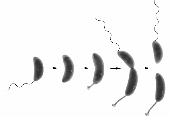
To begin with, I have to show you the canonical Caulobacter picture, from the lab of Yves Brun:

This is a picture that, for a while, seemed to be featured in every single paper and talk on the organism. It shows the “life cycle” of Caulobacter: it starts out (on the left) as a freely-swimming single cell. If it swims to a nice place, it sheds its flagellum, and grows that famous stalk and holdfast. Once anchored, it grows and divides, giving birth to a new free-swimming cell. Only the anchored cells divide, and once they’ve anchored, they never move again. So, as long as there is sufficient food, they’ll crank out swimming cells; when conditions turn bad, they are doomed.


Caulobacter, like a lot of bacteria, will form fairly dense growths of cells on a surface—a so-called biofilm. All the cells in a Caulobacter biofilm are growing on stalks, anchored by that marvelous glue—I can picture wandering through such a film, like wandering through a forest of oddly shaped trees that occasionally give birth to flagellated blobs. If conditions are good in this biofilm forest, then it’s advantageous for the newly-hatched Caulobacter swimmers to immediately stop swimming, grow a holdfast, and settle right in place—and so they do. The Caulobacter doesn’t fall far from the tree--sometimes they even cling to each other.


However, if times are tough, all the food has been used up and there are predators around, the existing Caulobacter might wish to urge their offspring to move on a bit before they settle down. A group of researchers, led by Yves Brun, found that this is exactly what happens: distressed Caulobacter biofilms produce something that essentially keeps Caulobacter glue from sticking.


There are several examples of Bacterial cells communicating with members of the same species using a variety of small molecules, so Brun set to work to figure out what Caulobacter used. Surprisingly, the molecule that inactivates Caulobacter glue is Caulobacter DNA. Brun found that a lot of cells in a stressed-out biofilm die, and shed their DNA into the area—and this DNA specifically interacts with the newly-formed holdfast of a former swimming cell, and completely turns off the glue.


Brun was commendably thorough in trying to prove that this was really happening. Unlike other known chemical signals used by Bacteria, the DNA didn’t trigger any changes in gene expression—so it wasn’t the case that the DNA stopped production of the glue: it inactivated the glue that was already there. His group purified large quantities of Caulobacter holdfasts, and found that the DNA only attached to the sticky end of the holdfast. The DNA didn’t interact with any other part of the cell. So, it seemed that the DNA from dead Caulobacter cells was a very specific “anti-glue.”


Here’s the really weird bit: only Caulobacter DNA was effective as an anti-glue. They tried DNA from other organisms; they tried DNA from distant and near relatives of Caulobacter—and only Caulobacter DNA was effective as an anti-glue! Some close relatives’ DNA could slightly reduce the glue’s effectiveness—but not as effectively as Caulobacter.


This is a real puzzle, and as yet there’s no satisfactory explanation. This is a glue that can stick to glass, plastic, rock, metal, you name it. Brun’s group tried DNA with the same overall chemical composition—the same percentage of G:C bases as Caulobacter—and got nothing. They searched the Caulobacter genome for distinctive genes or motifs—and got nothing. They looked for Caulobacter proteins or other small molecules contaminating their DNA—and got nothing. There is something yet to be found about Caulobacter DNA that does this.


There is something interesting in the fact that this signal that says “Warning! Go Away!” comes from dead cells.


We could look upon this as being a perfectly sensible system for free-swimming cells to avoid settling down where conditions are bad. The presence of loose Caulobacter DNA is a sure sign that Caulobacter cells have been there, and died. So, from the swimming cell’s point of view, it is not the place to settle down.


However, there is a growing number of researchers who look upon Bacteria as being social organisms—organisms that will not just communicate with each other, but also lay down their lives for others of the same species. So, these researchers say, look at it from the point of view of the stalked cell in the biofilm. You’re producing swimmer cells, each of which has 100% of your DNA. If conditions are bad, it would be worth your life to urge your offspring to get away. From this point of view, cells can actually commit suicide just to send that message.


This debate is as yet unresolved, and this recent finding about glue and antiglue doesn’t answer it one way or the other. However, it makes the amazing glue of Caulobacter seem even more amazing, and forces us to re-write the advertising copy: “Sticks like crazy to EVERYTHING!!!*”


(*except certain DNA sequences. We don’t know why.)

Berne, Cecile, David T. Kysela, and Yves V. Brun (2010). A Bacterial Extracellular DNA Inhibits Settling of Microbial Progeny Cells Within a Biofilm. Molecular Microbiology 77(4), 815-829.