
The other is a tease for next Friday:


A couple of days ago I showed off my students’ pictures of Caulobacter—the odd Bacterium that grows on a stalk; how that stalk is attached to a surface by a wonderful adhesive, which happens to be the strongest known natural glue. I remember when this finding was first publicized, I wondered—how do you measure how hard a single cell grabs on to its substrate? And, what sort of substance could a cell make to do this?
Alas, we still don’t know how to make Caulobacter glue. It seems to be made of some of the same material as the cell wall—a type of modified sugar polymer. Evidence of this is provided by the observation that a substance that weakens cell walls also weakens the Caulobacter glue. However, the glue is like a high-tech composite material: a lot of the sugar polymer doped with trace amounts of other chemicals to give it remarkable properties. We’re a long way from knowing all the ingredients, so don’t go looking for Caulobacter-brand glue anytime soon.
On the other hand, measuring the grip of a single cell is relatively straightforward. Essentially, all you need is a really small pair of tweezers and a leetle, tiny scale. The problem is getting fine enough tweezers to grab a single cell and a well-calibrated scale that can measure tenths of microNewtons—way less than a milligram.
People had tried this before. One old-school way is like the big bad wolf’s way of testing how well the little piggies’ houses were attached to their foundations: basically, put bacterial cells into a flow chamber, and see how fast a flow of water—and how much force—was required to blow them off their attachments.
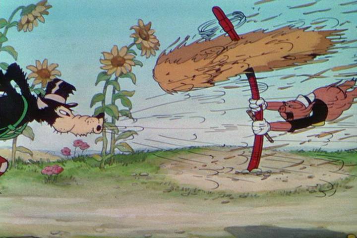 While this works for other bacteria, it simply didn’t provide enough force for blowing away Caulobacter. Furthermore, it only works on large populations. You can’t examine single cells this way.
A high-tech approach for measuring bacterial grip uses “optical tweezers,” which are essentially opposing laser beams. At the scale of bacterial cells, coherent light has substantial momentum, so a bacterial cell pinched between two lasers is experiencing something like a person immobilized by a pair of opposing fire hoses; you can then move the hapless victim around, and precisely measure the forces required (firemen actually have a game based on this principle). This works for moving and measuring the forces on run-of-the-mill Bacteria, but mighty Caulobacter can hold on so tight that optical tweezers won’t budge it.
Enter Peter Tsang and Ben Freund, using some rather crude technology to supply the brute force. They let Caulobacter attach to a thin cantilever of glass. They then seized the body of the Caulobacter cell with a powerful vacuum delivered by a micropipette—most of the cell was actually in the pipette, with just the stalk protruding. When they pulled on the pipette, the stalk stayed attached to the glass—but the glass was bent by the force. Here’s a cartoon version of the set-up:

It’s kind of a far-fetched image, but imagine Caulobacter was a person who had glued his feet to a diving board—Tsang and Freund grabbed on to his head with a giant vacuum cleaner, and pulled up. The harder they pulled, the more the diving board was bent up—and you can precisely measure the force required to move the diving board, which allows you to estimate how strong the glue is. If you know the shoe size of the guy on the diving board, you can say that the glue can withstand so many kilograms of force per square centimeter. This movie shows the microscope’s-eye view of the experiment:
At first, we see the pipette grabbing on to a single Cauobacter cell, one of many on the thin piece of glass. As more and more force is applied, the glass moves further and further, until—TWANG!—something breaks.
From there, it’s a fairly simple matter to figure out the force required; knowing how big the footprint of a Caulobacter cell is allows us to figure out the strength of the glue.
Actually, what we really know is a lower limit for the strength of the glue. When the researchers took a really close look at what happened, they found that every time, the glue didn’t fail—the stem itself snapped. So, the glue is actually stronger than what they calculate. The previous record holder for “best natural adhesive” were the microscopic bristles of gecko feet (themselves a neat story), which can hold about a kilogram per square millimeter. The glue holding my dental crown in place can hold about 3 kilos per square millimeter. The best we know about Caulobacter glue—whatever it’s made of—is that it can hold at least 6.8 kilos per square millimeter.
This investigation was really as much about engineering as biology—the kind of thing that is really just neat, but could have some practical value at some distant date. For Caulobacter, though, it’s just part of living. And, it turns out, there’s more to the story…


 Of course, my Mom's garden is cooler than mine. We have a good crop of mud right now.
There’s a lot we don’t know about GTAs—and there’s lots of evidence that they may be hugely important in the evolution of many Bacterial and Archaeal lineages. A report from October of 2010 finally provides some solid clues to just how important GTAs are for “wild” cells. Up to this time, GTAs had been either studied in the lab, or merely predicted from analysis of genome data. A group led by John H. Paul at the University of Florida decided to see if GTAs actually worked in “wild” cells. This required a little genetic manipulation—so the study is not absolutely pure—but their results show the importance of GTAs in natural systems.
The first thing that Paul’s group did was to find “wild” cells that could produce GTAs. They added a few easily detected genes to these cells—specifically, genes for antibiotic resistance. If GTAs actually moved genes from one cell to another, then GTAs from these cells could make “recipient” cells resistant to antibiotics, and thus very easy to find. However, they wanted to see if GTAs worked in the wild. So, they mixed together GTAs from their donor cells and samples of seawater in the natural environment. The photo shows their experimental apparatus, which contained the cells so that no GMOs would be released into the environment.

After a day, they checked to see if any of the cells in their seawater sample became resistant to antibiotics. Sure enough, many did—as many as one in 100 cells.
This looks like proof that GTAs could transfer genes “in the wild.” However, the researchers still needed to eliminate an alternative hypothesis—that the cells in their seawater spontaneously became resistant to antibiotics. This did actually happen, but at a much, much lower rate—when they tried the same experiment without any GTAs, they found that 1 in 10,000 cells spontaneously became antibiotic resistant.
This finding forces us to re-evaluate how often genes get shared among different organisms in the real world. Paul’s group found that GTAs could efficiently transfer genes between fairly dissimilar types of bacteria. This is something viruses are not so good at (and it's also the reason that you don’t have to worry about catching a viral infection from frogs). Also, they found that the efficiency of gene transfer by GTAs is about a million times that of transduction. So, in a very real sense, the ocean is swimming with genes, and species evolve influenced not just by the physical parameters of temperature and light, but also by the genes that are there for the taking. Genomes are far more flexible than we previously thought, constantly acquiring new genes and trying them out, and evolving with astonishing speed. Evolution is not an orderly, linear march; it is a spastic tango, darting and weaving and messily exchanging genes all over the place.
Lang, Andrew S, and J. Thomas Beatty (2006). Importance of widespread gene transfer agent genes in a-proteobacteria. TRENDS in Microbiology 15: 2, 54-62.
Marrs, Barry (1974). Genetic Recombination in Rhodopseudomonas capsulata. Proceedings National Academy of Science USA 71:3, 971-973.
McDaniel, Lauren, Elizabeth Young, Jennifer Delaney, Fabian Ruhnau,
Kim B. Ritchie, and John H. Paul (2010). High Frequency of Horizontal Gene Transfer in the Oceans. Science 330, 50.
Solioz, Marc, Huei-Che Yen, and Barry Marrs (1975). Release and Uptake of Gene Transfer Agent by Rhodopseudomonas capsulata. Journal of Bacteriology 123:2, 651-657.
Yen, H.C., N. T. Hu, and Barry L. Marrs (1979) Characterization of the Gene Transfer Agent Made by an Overproducer Mutant of Rhodopseudomonas capsulata. Journal of Molecular Biology 131, 157-168.

(Click here for part I, an introduction to transduction)
There is a twist on transduction, using viruses to transfer genes between cells. It’s a kink that was first discovered in 1974, but it still is not well understood. Barry Marrs of Saint Louis University was analyzing strains of a bacterium called Rhodobacter, and found that certain strains of this organism were capable of transferring genes to other strains with surprising efficiency. In some ways, Marrs’ discovery seemed very similar to transduction: something involving DNA in protein envelopes was involved, and random pieces of the cells’ genome were carried in these envelopes.
Despite the superficial similarity to transduction, it was apparent that something unique was going on. Transduction is a rare event—for every transducing virus produced, a hundred thousand killer viruses are made. However, Marrs and others observed that the gene transfer was never associated with any lethality; it was as if the viruses were only carrying cellular DNA, never viral DNA. Other differences were noticed as well. The “gene transfer agents,” or GTAs as Marrs called them, were far smaller than any known virus. Although they could be observed by electron microscopy, and had the same shape as a regular virus, they were relatively tiny; here is a transmission electron micrograph of purified GTAs, showing their resemblance to tailed viruses.

A normal virus could hold a few dozen genes, but the GTAs could only carry four or five at a time. This small size makes it clear that whatever GTAs may be, they are not viruses. Remember that a virus particle is a protein envelope carrying the genes for making more virus particles—so the DNA encoding the virus must fit within a virus particle. We know the genes required for making GTAs, and how long a piece of DNA that requires—and it is impossible for the DNA encoding GTA production to actually fit in a GTA particle! It's as if the instructions for making a box were far, far larger than the box itself.
So what are GTAs? This is still an open question. We know from experiments and the analysis of genomes that there are many different types of GTAs, and they are found in Bacteria and Archaea. They always “look” like viruses. The genes encoding GTAs are organized in a pattern reminiscent of some viruses, and some of the individual genes for GTAs are homologous to viral genes--but there's always the problem that there's no way to fit all the genes for a GTA into a GTA.
However, while the execution of the construction program for many viruses has been dissected in excruciating detail, we know next to nothing about how GTAs are actually produced. We don’t even know such basic facts as whether, as with viruses, GTA production results in the destruction of the “donor” cell. The presence of a gene similar to viral “lysis” genes in many GTA gene clusters is suggestive—but it is not a smoking gun, and there's no picture like this

of GTAs escaping a cell. All we really know is that GTA genes are under cellular control, and are typically only expressed when the cells are crowded—conditions which make it more likely for a GTA to find a susceptible target.
The evolutionary history of GTAs is also wrapped in mystery. Their homology to viruses is suggestive of some sort of shared ancestry, but the nature of the connection is obscure. One can make a "family tree" of the proteins in viruses, and you'll see that a handful of GTA proteins fit very nicely in that tree. You can also make a family tree of the proteins in GTAs, and a handful of viral proteins fit right in. So, the only thing we can say is clear is that the viruses and GTAs have some shared family history--but who is descended from whom?
Are GTAs viruses that have been domesticated? Perhaps. The widespread distribution of viruses throughout the “tree of life,” and the relative rarity of GTAs has been used as an argument for this case. In this view, a twig on the family tree of viruses mutated to become GTAs, and they have been preserved for their role in facilitating genetic exchange. On the other hand, there are certain viruses that have genomes containing genes commonly found in viruses as well as genes that are usually used in making GTAs. The earliest reports of GTAs argue that gene transfer is as necessary to Bacteria and Archaea as sex is to eukaryotes. In this view, GTAs predate viruses; viruses are GTAs that "went rogue."
There is another aspect to GTA evolution that is puzzling. Although it is not known for certain, the similarities to viruses suggest that GTA production is fatal. The GTA genes thus serve no benefit to the individual cell—in fact, they are absolutely detrimental. Traditional views of natural selection imply that such genes should not be preserved—to this view, it is every bacterium for itself and damn the rest. The notion that a single bacterial cell should commit suicide and share its genes with its neighbors for their benefit is anathema. However, GTAs may be evidence of “kin selection,” where a gene benefits the species at the expense of the individual. The persistence of GTA genes in many different microbial lineages suggests that this may indeed be the case.
Such a view is heresy--I constantly remind my students that genes work for the good of the individual, never for the good of the species. However, GTAs may force me to put an asterisk by that statement and rethink evolution, or (more likely) redefine "individual" to include "a group of genetically identical yet independently living cells."


 It got down to -3 degrees C Sunday night. Click on the picture to see the dinos-hoar.
Viruses play a huge role in the lives of microbes. We are used to thinking of viruses in human terms, and for humans—whether it’s a case of the sniffles caused by rhinovirus, or death from poxvirus—viruses are agents of disease. But, for the Bacteria and Archaea, viruses are much more. They are agents of evolution.
Structurally, a virus is much simpler than a cell. It is essentially an envelope of made of protein, carrying a bunch of genes on a piece of DNA or RNA. The protein envelope is supremely effective at attaching to the surface of a specific type of cell, and once it has done so, it injects the DNA or RNA it carries into that cell. Once inside the cell, the virus’ genes will instruct the cell to make lots of those envelope proteins and make lots of copies of the genes, and assemble them into new viruses. Usually, the virus genes also encode a protein that pops the cell open, killing the cell and releasing hundreds of new viruses so the cycle can start again. Most of the time—like, 99.9999% of the time—this is what happens when a virus attacks a Bacterial or Archaeal cell. However, what happens the other .00001% of the time is hugely important for the evolution of these cells.
I had an awful nightmare once—too much studying!—one of those horrible dreams where you’re in two places at once, and neither is nice. In this dream, I was a citizen of Berlin as it was overrun by the Russian army at the end of WWII...
and I was also a ribosome in a bacterial cell that was being attacked by a virus. Everything was chaos and confusion and terror. Easy enough to see in the Berliners; the second picture shows a cell exploding after viral infection. Each little pale tadpole-shaped thing is a new virus particle; remember that shape.
In such a scene it’s no surprise that mistakes are made, and when a bacterial cell dies at the microscopic hands of a virus, not everything goes according to virus’ program. Every once in a while, by random chance, a piece of the bacterial cell’s DNA ends up in a virus envelope, instead of viral DNA.
This “virus with bacterial genes” can go and attach itself to another bacterial cell and inject its DNA—but instead of that cell getting sick, it will get some perfectly good (if second-hand) bacterial genes. These genes could potentially increase the fitness of the cell, and so they could change the course of evolution.
This process is called “transduction,” and it is a major way in which Bacteria and Archaea can exchange genes. For humans (and all eukaryotes), sex is a medium for genetic exchange, and it can affect the course of evolution by putting genes into new and potentially useful combinations. The Bacteria and Archaea don’t have sex, but they do have transduction to shuffle the genetic cards (even though the DNA donor dies in the process). As one of my professors joked, in humans, sex brings viruses; but in the Bacteria and Archaea, viruses bring sex.
Transduction is well known and described. It has even been “domesticated” by humans so it is a commonly used tool in microbial genetics research. If I wished to move a gene from one bacterium to another, I’d grow up a trillion of my “donor” bacteria, infect them with viruses, wait a little bit, and collect the new viruses that were produced. I’d then go and take these viruses and infect a trillion of my “recipient” cells with them. It would be a massacre—all of the donor cells, and almost every single one of those recipient cells would die. But some would be lucky, infected by a transducing virus instead of a normal virus, and some small number of those would be even luckier—they would have the gene that I wanted to move.
Transduction is statistically rare—but given the numbers of microbes in the environment, transduction is also amazingly common. A milliliter of seawater contains some 1,000,000 bacteria and perhaps some 10,000,000 bacterial viruses. Plug in the number of milliliters in all the oceans and there are probably well over 1030 viruses swimming around out there, or as Ken Stedman put it, the same mass as 75 million blue whales. Given what we know about the frequency of transduction, this sort of bacterial sex happens, oh, about 20,000,000,000,000,000 times every second.
Think about that next time you dip your toes in the ocean.
(Sources--any reputable micro textbook, Ken Stedman of Portland State University, U.S. Army Museum)
See, you majors in microbiology,
students of small,
Diverse as the peoples of earth!
You would know
The range of microbes,
More than the stars of heaven!
Beijerinck taught us,
Kluyver taught us,
Van Niel taught us,
Hungate taught us,
Stainer taught us,
Woese and Wolfe taught us,
And now we must teach you.
See! How many the microbes,
They crowd the earth
Like stars of heaven.
Understand them,
Count their number,
And call them all by name!
