Showing posts with label cell structure. Show all posts

Thursday, January 27, 2011

Caulobacter Crazy Glue

A couple of days ago I showed off my students’ pictures of Caulobacter—the odd Bacterium that grows on a stalk; how that stalk is attached to a surface by a wonderful adhesive, which happens to be the strongest known natural glue. I remember when this finding was first publicized, I wondered—how do you measure how hard a single cell grabs on to its substrate? And, what sort of substance could a cell make to do this?


Alas, we still don’t know how to make Caulobacter glue. It seems to be made of some of the same material as the cell wall—a type of modified sugar polymer. Evidence of this is provided by the observation that a substance that weakens cell walls also weakens the Caulobacter glue. However, the glue is like a high-tech composite material: a lot of the sugar polymer doped with trace amounts of other chemicals to give it remarkable properties. We’re a long way from knowing all the ingredients, so don’t go looking for Caulobacter-brand glue anytime soon.


On the other hand, measuring the grip of a single cell is relatively straightforward. Essentially, all you need is a really small pair of tweezers and a leetle, tiny scale. The problem is getting fine enough tweezers to grab a single cell and a well-calibrated scale that can measure tenths of microNewtons—way less than a milligram.


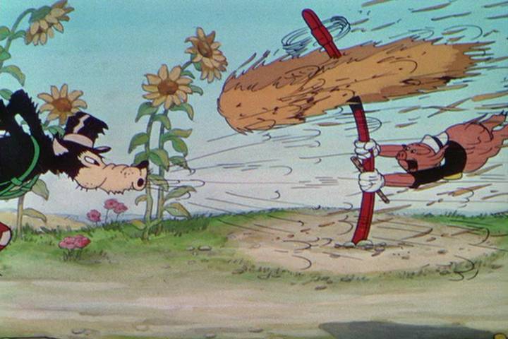
People had tried this before. One old-school way is like the big bad wolf’s way of testing how well the little piggies’ houses were attached to their foundations: basically, put bacterial cells into a flow chamber, and see how fast a flow of water—and how much force—was required to blow them off their attachments.

While this works for other bacteria, it simply didn’t provide enough force for blowing away Caulobacter. Furthermore, it only works on large populations. You can’t examine single cells this way.


A high-tech approach for measuring bacterial grip uses “optical tweezers,” which are essentially opposing laser beams. At the scale of bacterial cells, coherent light has substantial momentum, so a bacterial cell pinched between two lasers is experiencing something like a person immobilized by a pair of opposing fire hoses; you can then move the hapless victim around, and precisely measure the forces required (firemen actually have a game based on this principle). This works for moving and measuring the forces on run-of-the-mill Bacteria, but mighty Caulobacter can hold on so tight that optical tweezers won’t budge it.


Enter Peter Tsang and Ben Freund, using some rather crude technology to supply the brute force. They let Caulobacter attach to a thin cantilever of glass. They then seized the body of the Caulobacter cell with a powerful vacuum delivered by a micropipette—most of the cell was actually in the pipette, with just the stalk protruding. When they pulled on the pipette, the stalk stayed attached to the glass—but the glass was bent by the force. Here’s a cartoon version of the set-up:

It’s kind of a far-fetched image, but imagine Caulobacter was a person who had glued his feet to a diving board—Tsang and Freund grabbed on to his head with a giant vacuum cleaner, and pulled up. The harder they pulled, the more the diving board was bent up—and you can precisely measure the force required to move the diving board, which allows you to estimate how strong the glue is. If you know the shoe size of the guy on the diving board, you can say that the glue can withstand so many kilograms of force per square centimeter. This movie shows the microscope’s-eye view of the experiment:

At first, we see the pipette grabbing on to a single Cauobacter cell, one of many on the thin piece of glass. As more and more force is applied, the glass moves further and further, until—TWANG!—something breaks.


From there, it’s a fairly simple matter to figure out the force required; knowing how big the footprint of a Caulobacter cell is allows us to figure out the strength of the glue.


Actually, what we really know is a lower limit for the strength of the glue. When the researchers took a really close look at what happened, they found that every time, the glue didn’t fail—the stem itself snapped. So, the glue is actually stronger than what they calculate. The previous record holder for “best natural adhesive” were the microscopic bristles of gecko feet (themselves a neat story), which can hold about a kilogram per square millimeter. The glue holding my dental crown in place can hold about 3 kilos per square millimeter. The best we know about Caulobacter glue—whatever it’s made of—is that it can hold at least 6.8 kilos per square millimeter.


This investigation was really as much about engineering as biology—the kind of thing that is really just neat, but could have some practical value at some distant date. For Caulobacter, though, it’s just part of living. And, it turns out, there’s more to the story…


Peter Tsang, Guanglai Li, Yves Brun, L. Ben Freund, and Jay X. Tang (2006). Adhesion of Single Bacterial Cells in the Micronewton Range. Proceedings of the National Academy of Science USA 103(15):5764-8.

Tuesday, May 11, 2010

Insane in the (Outer) membrane

Okay, this is weird, even by Archaeal standards. That’s like saying that an outfit is eccentric, even by Lady Gaga standards. But this recent discovery about the structure of an unusual Archaeal cell is just a bit much: it takes one of the most basic features of cell structure and turns it inside out.

The Archaea are a group of organisms with small cells that are superficially similar to Bacteria. However, the genetic and biochemical differences between Bacteria and Archaea are as great as the differences between Bacteria and us, the Eukaryotes. Archaea have earned a reputation as oddballs. Many are “extremophiles,” living in environments with paint-peeling pH or scalding temperatures or pickling concentrations of brine. But in spite of all the differences between the Eukaryotes, Bacteria and Archaea, we are all descended from a common pool of ancestors, which accounts for our similarities: we all have inherited the same genetic coding and decoding system, and use similar mechanisms for getting energy.


This may look like a picture of Hoover Dam, but it’s actually a graphic explaining how almost every cell gets energy. There’s too much water upstream of the dam; there’s not enough water downstream of the dam. If you let water go through the dam, energy is released that you can use to do work—in this case, turn electrical generators. The system keeps recharging itself, as energy from the environment (the sun) does the work of lifting water back up into the atmosphere, eventually filling the river that feeds the dam.

This is how cells make energy. The cell’s membrane acts as a dam. Outside the cell, there are too many positive ions, while inside the cell there aren’t enough. If the cell lets positive ions in, energy is released that can do work. In this case, the energy drives a protein called ATPase that sits in the membrane. ATPase makes ATP, the chemical that powers just about everything in the cell. The cell keeps recharging itself by taking energy from the environment (sunlight or food) to push positive ions back out of the cell and “refilling” the reservoir (the outside of the cell).

The power from this “energized” membrane is used not only to make ATP, but also to move stuff into and out of the cell. There are a few cells that make ATP by a different mechanism, fermentation; but they still use much of their ATP to energize their membranes. An energized membrane is one of the hallmarks of any living cell, Bacterial, Eukaryotic, or Archaeal.
This universal mechanism for storing energy shows up in the architecture of every cell. In Bacteria, it’s like so:

The inner membrane is the energized membrane, where the ATPase is found. The ATP that is produced diffuses into the cell, where it is used to copy DNA and make proteins and power all the essential work of the cell. The space between the inner and outer membranes is small, less than 20% of the cell’s volume; it has a relatively high concentration of positive ions, and no ATP is found there.

Eukaryotes have mitochondria to make most of their ATP. The mitochondrion is actually evolved from bacteria that took up residence in the Eukaryotic cell, so we see basically the same arrangement of membranes and ATPase. The only difference is that special pores move ATP out of the mitochondrion and into the rest of the cell:


Archaeal cells are pretty much the same as Bacterial cells, except there’s no outer membrane. In every case, Bacterial, Eukaryotic, or Archaeal, the inner membrane is the energized membrane—the cell’s Hoover Dam.


Researchers led by Harald Huber of Regensberg, Germany, have been patiently studying the Archaean Ignicoccus hospitalis. This organism was found growing in near-boiling water at a geothermal vent in the Midatlantic Ridge, but it has been domesticated and grown in the laboratory in a mixture of hydrogen, elemental sulfur, and carbon dioxide at a temperature of 90 degrees Celsius. This passes for normal behavior in Archaea. The first surprise came in 2002 when Huber’s group used an electron microscope to examine these cells. What we’re looking at here is a very thin slice of a cell.


Now this is weird. There are clearly two membranes, and no cell wall. Bacteria generally have two membranes, but the Archea? No. And the space between the inner and outer membranes? HUGE! It’s 75% of the cell’s volume! In the Bacteria, that compartment is at most 20% of the cell’s volume. All the business of the cell—all the DNA, all the gene expression, all the synthesis of macromolecules and so on—goes on in the darker region, inside what we’ll call the inner membrane.

This bizarre structure raises questions. Since Ignicoccus’ structure resembles that of a Bacterium, Huber's group assumed it would be organized like a Bacterium, with an energized inner membrane. But why would so much of the cell—all that space between the membranes—not get any energy? So, they decided to look for ATPase. Since ATPase is too small to show up even in an electron microscope, they used a chemical trick to “tag” each molecule of ATPase with a little blob of gold. This shows up as a little black dot under an electron microscope. Here’s what they found; each black dot is a molecule of ATPase with a gold-tagged antibody stuck to it:


This picture makes as much sense to me as this:

I think that Huber’s group thought the same, so they checked and re-checked their result by a couple of completely different methods. The result was confirmed, and is baffling. The outer membrane is full of ATPase; the space between the membranes must get plenty of ATP. But all the DNA, all the gene expression, all the protein synthesis—all the stuff that consumes ATP—is going on inside the inner membrane. Nothing like a normal Archaeal cell, and nothing like a normal Bacterial cell.

The last page of the article describing this discovery is basically a long list of questions raised by this weird structure. Some of these questions are mechanical: if all the DNA and gene expression machinery is inside the inner membrane, how does ATP get through the inner membrane? What goes on in the space between membranes? Is the inner membrane energized? Even more puzzling are the evolutionary questions raised by Ignicoccus’ structure and the location of its ATPase. Ignicoccus is the only cell we know—perhaps in the entire world—with an energized outer membrane. Since all the other Archaea have just one membrane, it’s likely that Ignicoccus evolved from something with just one membrane. How did that happen? Is the outer membrane the recent addition—and if so, how did ATPase end up there? Is the inner membrane the recent addition—and if so, how did it evolve a mechanism to import ATP?

On the one hand, this discovery is not terribly significant; Ignicoccus is a niche player in a highly restricted environment, and it has no close relatives. On the other hand, it raises a lot of big questions about how such a freakish cell could evolve. These questions are relevant to us. A leading hypothesis for the origin of Eukaryotic cells posits that an Archaeal cell engulfed a bacterial cell; the Bacterium became the mitochondrion, while the Archaeal DNA was protected by an internal membrane. Huber notes that an Archaeal cell similar to Ignicoccus could be our great-great-to-the-gazillion-grandaddy.

Ulf Küpera, Carolin Meyerb, Volker Müllerc, Reinhard Rachelb, and Harald Hubera (2009). Energized outer membrane and spatial separation of metabolic processes in the hyperthermophilic Archaeon Ignicoccus hospitalis. PNAS 107:7, 3152-3156.

REINHARD RACHEL, IRITH WYSCHKONY, SABINE RIEHL and HARALD HUBER (2001). The ultrastructure of Ignicoccus: Evidence for a novel outer membrane and for intracellular vesicle budding in an archaeon. Archaea 1, 9-18.

Friday, May 7, 2010

Bacterial neat freaks

The first time I taught a college biology class, I told my students about the structures of different types of cells. There was our type of cell, the eukaryotic cell, with its nucleus like an egg yolk, a web of cytoskeleton, and the collection of mitochondria, endoplasmic reticulum, Golgi apparatus, vesicles, and other “organelles”. All these things make the eukaryotic cell an orderly and structured place. In contrast, there was the Bacterial cell, which I presented as a featureless bag of enzymes. Nobody at the time had convincingly described any distinctive structure or compartments within a bacterial cell. Organelles and cytoskeletons, I told my students, were strictly eukaryotic features.
I always caution my students that I am going to teach them some stuff that is simply wrong. We have recently learned that most Bacteria have a cytoskeleton, and that it shares its ancestry with the eukaryotic cytoskeleton. We are also finding out that there are several different types of organelles within Bacterial cells. A recent study from Pam Silver’s group shows just how sophisticated Bacterial cell structure can be.


Silver’s group studied a type of Bacterium that does photosynthesis: it uses light to make energy (“photo”) and then takes hydrogen from water and adds it to carbon dioxide to synthesize sugar (“synthesis”).


These bacteria are cylindrical, roughly the shape of a tennis-ball can. The “photo” part of their photosynthesis takes part on the surface of the cell, but the “synthesis” part happens in the cell’s interior. The enzyme that is responsible for starting synthesis, called Rubisco, is not efficient. It is like a factory worker with a specific job to do on an assembly line, such as putting together two widgets to make a bigger widget. But this worker is clumsy about picking up the widgets from his workplace, and once he has them, he’s slow to put them together. Rubisco puts together two widgets: carbon dioxide, and a type of sugar, making a bigger sugar molecule. But Rubisco is bad at picking up carbon dioxide, and once it has it, it’s slow to use it.

If you were the sluggish worker’s foreman, you could try to improve his efficiency by putting that worker in a small room. If you filled that room from floor to ceiling with lots of the widgets he needed, the only thing he could pick up would be work for him to do. If you kept all the other different types of workers and their tools and widgets away, he wouldn’t get distracted. Bacterial cells make these small rooms; they are called carboxysomes, and they are one of the best-studied examples of bacterial organelles.

The carboxysome is a room in the bacterial cell reserved for Rubisco. If the cell is the size of our can of tennis balls, the carboxysome is about the size of a ping-pong ball. The room has a few thousand molecules of Rubisco gluing together carbon dioxide and sugar. The walls of this room are particularly remarkable. They are made of a protein called “Ccm K4”. Ccm stands for “carbon-dioxide concentrating membrane,” and it describes precisely what this protein does: the only things that can enter the carboxysome are sugar and carbon dioxide. Everything that Rubisco sees is work for it to do. Without the carboxysome (for example if the cell can’t make Ccm K4 protein), rubisco is so inefficient that photosynthesis grinds to a halt.
Silver’s group wanted to show that the carboxysome is made of CcmK4 and full of rubisco. So, they made a version of the Ccm K4 protein that glowed red, and a version of the Rubisco protein that glowed green. Looking under the microscope, they saw that the cells they were studying had blobs of the right size, glowing both red and green. This was nice but not particularly surprising, since something similar had been seen in other Bacteria. However, they were surprised to see that the carboxysomes were all precisely lined up, right along the long axis of the cell, and that they were very evenly spaced.
If the bacterial cell were the unstructured “bag of enzymes” that I used to lecture about, then a small object like a carboxysome should move around within the cell by diffusion—the same process that makes a drop of ink disperse in a cup of water. The random motion of molecules would push carboxysomes around, and if there were more than one carboxysome per cell, they should be jumbled around. But the carboxysomes did not move at all: they stayed in their evenly spaced order.

Comparison of these cells with other bacterial cells suggested that they have a cytoskeleton: filaments of proteins that run the length of the cell. There are two different types of protein that make up the cytoskeleton in these cells. When Silver’s group made cells that didn’t have one of these proteins, the cells lost their cylindrical shape; instead of looking like a can of tennis balls, the cells looked like balloons. This protein acted like a spring, pushing out the ends of the cell to make it cylindrical. Their carboxysomes were randomly distributed throughout the cell.

When Silver’s group removed the second type of cytoskeletal protein, called “Par A,” from the cell, they saw something really interesting. The cells were still cylindrical, so the bulk of the cytoskeleton was still present. However, the carboxysomes were jumbled around: in some cells, they were clumped together in the middle, in others, there was a pile of carboxysomes at one end or the other. The Par A protein somehow aligns and spaces the carboxysomes.
Par A is a very busy protein; each Par A molecule spends energy moving itself from one end of the cell to the other, then back again; it’s as if Par A has no purpose but to wander back and forth. The cell makes thousands of molecules of Par A, and all these molecules move essentially in unison, like a crowd of people shuffling en masse from one end of a hall to the other, with a round trip taking about two hours. Par A proteins have been seen in many other rod-shaped bacteria without carboxysomes, but Silver’s group was surprised to see that there was a lot of Par A protein oscillating between each of the carboxysomes in a cell. If Par A molecules get stuck between carboxysomes, they still wander back and forth, but over a much shorter distance. The Par A that Silver’s group observed ping-ponging between carboxysomes is enough to nudge the carboxysomes apart. In the time it takes for most Par A molecules to make a round trip of the length of the cell, enough accumulates between carboxysomes to make them evenly spaced.

Why should a cell spend so much energy to arrange the carboxysomes so precisely? Silver’s group addressed this question by studying the cells that didn’t have Par A protein. Because they had a random arrangement of carboxysomes, when the cell divided there was a significant chance that one of the resulting cells would not have any carboxysomes. These cells could eventually make new carboxysomes, but they would grow much more slowly. So, one generation’s energy investment in making organelles and arranging them pays off in the next generation.

There’s at least one thing that’s much more important to the survival of the cell than a carboxysome—the cell’s DNA. It turns out that a Par A system can be used to push one copy of the cell’s DNA to each end of the cell before the cell divides. The name “Par A” is actually short for “DNA partitioning protein A”. Silver speculates that the mechanism for arranging carboxysomes evolved from the DNA partitioning system, since they both make sure that each of the products of cell division gets a necessary feature. What’s more, this Bacterial Par A protein is similar to the protein, actin, that makes up much of the cytoskeleton in Eukaryotic cells. Some sort of cytoskeleton was probably a feature in the last common ancestor of all living things.

So, if any of my 1998 students are reading this, please revise what I told you about Bacteria. They are not just bags of enzymes. Many have cytoskeletons and organelles—they’re even more like you and me than we thought.

David F. Savage, Bruno Afonso, Anna H. Chen, Pamela A. Silver (2010). Spatially Ordered Dynamics of the Bacterial Carbon Fixation Machinery. Science 327, 1258-1261.